
NIH 3D
Chemokine receptor 2 (CCR2)

- Creator (as listed at the source)
- Not listed at the source.
- Licence
- Public DomainStated at the source marketplace; SliSearch does not verify licences.
- File types
- GLB STL
- Pricing
- Free
- Published
- Category
- Biomacromolecules
- Downloads
- 58
Overview
a 3d model generated using pymol of chemokine receptor 2 (CCR2)
Print this with Prinnu
- Download the model file from the source page. SliSearch moves no files.
- Upload the file at slicience.com/quote. Prinnu quotes it in minutes.
Similar models
Chosen by shared tags first, then category. Each one links to its own marketplace.
 The 2.5 A structure of the CXCR4 chemokine receptor in complex with small molecule antagonist IT1t
 Progesterone Receptor bound to DNA
 The structure of UNC5b cytoplasmic domainPDB Classifiation: APOPTOSIS
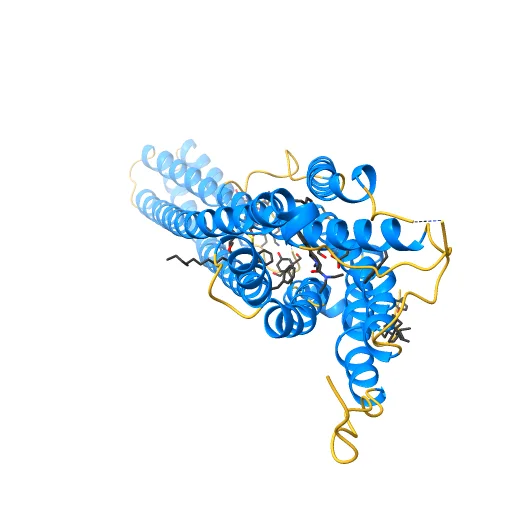 Crystal structure of the LSD-bound 5-HT2B receptor
 Crystal Structure of the Kinase domain of Human HER2 (erbB2).PDB Classifiation: TRANSFERASE/TRANSFERASE INHIBITOR
 Crystal structure of BcTSPO, type 2 at 1.7 Angstrom with DMSOPDB Classifiation: MEMBRANE PROTEIN
 Insulin Receptor ectodomain in complex with two insulin molecules
 Crystal structure of the human TLR4-human MD-2-E.coli LPS Ra complexPDB Classifiation: IMMUNE SYSTEM
 Structure of the TRPM8 cold receptor by single particle electron cryo-microscopy, calcium-bound stateTRPM8 cold receptor, calcium-bound state
 Structure of the TRPA1 ion channel determined by electron cryo-microscopyPDB Classifiation: TRANSPORT PROTEIN
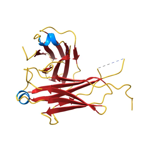 IgM C2-domain from mousePDB Classifiation: PROTEIN BINDING
 FERRIC ENTEROBACTIN RECEPTORPDB Classifiation: MEMBRANE PROTEIN
 Rhodopsin-Transducin Complex
 First and second Ig domains from human Robo1 (Form 2)PDB Classifiation: RECEPTOR
 Crystal Structure of a 1:1:1 FGF23-FGFR1c-aKlotho Ternary ComplexPDB Classifiation: HYDROLASE/PROTEIN BINDING
 Crystal structure of 5-HT2AR in complex with zotepinePDB Classifiation: MEMBRANE PROTEIN
 Diabody 330 complex with EpoRPDB Classifiation: protein binding/immune system
 Structure of the human histamine H1 receptor in complex with doxepin
 Electron Cryo-Microscopy of Chikungunya in Complex with Mouse Mxra8 ReceptorChikungunya in Complex with Mouse Mxra8 Receptor
 Cannabinoid Receptor 1
 HemagglutininThe 'H' in H1N1 (and all forms of influenza). This variant is based on the PDB structure 1RUZ from the 1918 pandemic, and is colored as follows to indicate sites of mutations between different variants.
 Crystal structure of the TLR1-TLR2 heterodimer induced by binding of a tri-acylated lipopeptidePDB Classifiation: IMMUNE SYSTEM
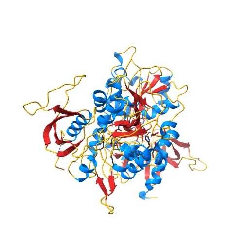 CRYSTAL STRUCTURE OF GLUN1/GLUN2A LIGAND-BINDING DOMAIN IN COMPLEX WITH GLYCINE AND NVP-AAM077PDB Classifiation: TRANSPORT PROTEIN
 CRYSTAL STRUCTURE OF THE ECTODOMAIN OF HUMAN TRANSFERRIN RECEPTOR
 Crystal Structure of a new rexinoid bound to the RXRalpha ligand binding doamin in the RXRalpha/PPARgamma heterodimerPDB Classifiation: HORMONE/GROWTH FACTOR
 PHOSPHORYLATED INSULIN RECEPTOR TYROSINE KINASE IN COMPLEX WITH PEPTIDE SUBSTRATE AND ATP ANALOG
 Cryo-EM structure of the human growth hormone-releasing hormone receptor-Gs protein complex
 Serotonin receptor
 Structure of the IL-10R2 Common ChainPDB Classifiation: PROTEIN BINDING
 MURINE T-CELL ANTIGEN RECEPTOR 2C CLONEPDB Classifiation: RECEPTOR
 Crystal structure of the human beta2 adrenoceptorPDB Classifiation: SIGNALING PROTEIN
 Structure of novel coronavirus spike receptor-binding domain complexed with its receptor ACE2PDB Classifiation: VIRAL PROTEIN
 Human GRK2 in Complex with Gbetagamma subunitsPDB Classifiation: Transferase/Signaling protein
 Crystal structure of VEGFR1 in complex with N-(4-Chlorophenyl)-2-((pyridin-4-ylmethyl)amino)benzamidePDB Classifiation: TRANSFERASE
 CB1 Receptor
 Acetylcholine binding protein (AChBP) as template for hierarchical in silico screening procedures to identify structurally novel ligands for the nicotinic receptorsPDB Classifiation: RECEPTOR
 Ligand Recognition by A-Class Eph Receptors: Crystal Structures of the EphA2 Ligand-Binding Domain and the EphA2/ephrin-A1 ComplexPDB Classifiation: TRANSFERASE/SIGNALING PROTEIN
 Crystal structure of the IL4-IL4R-Common Gamma ternary complexPDB Classifiation: Cytokine/Cytokine receptor
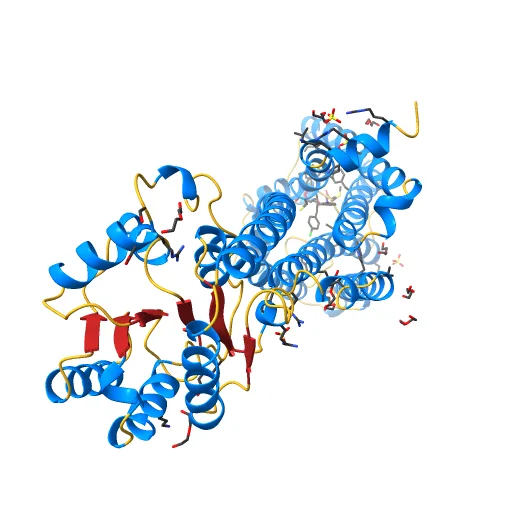 High-resolution crystal structure of the human CB1 cannabinoid receptor
 Apo form metabotropic glutamate receptor 5 with Nanobody 43Apo form metabotropic glutamate receptor 5 with Nanobody 43, primary map
SliSearch is an index: it stores public metadata only, does not host files, does not verify licences, and has no partnership or affiliation with the indexed marketplaces.