
NIH 3D
Crystal structure of the LSD-bound 5-HT2B receptor


- Creator (as listed at the source)
- Not listed at the source.
- Licence
- Public DomainThe licence is listed by the source marketplace. SliSearch does not verify it.
- File types
- GLB STL
- Pricing
- Free
- Published
- Category
- Biomacromolecules
- Downloads
- 226
Overview
The creator did not write a description for this model.
Print this model with Slicience
- Download the model from the source page.
- Upload the file to Slicience and get a quote with Prinnu.
Similar models
Chosen by shared tags first, then category. Each one links to its own marketplace.
 Crystal structure of Smad4-MH1 bound to the GGCGC site
 Crystal Structure of GDP-bound Human KRas
 CRYSTAL STRUCTURE OF THE ECTODOMAIN OF HUMAN TRANSFERRIN RECEPTOR
 Crystal structure of MtISWI bound with histone H4 tail
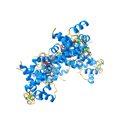 Crystal structure of endothelial nitric oxide synthase peptide bound to calmodulin
 Crystal structure of Aa-Ago with externally-bound siRNA
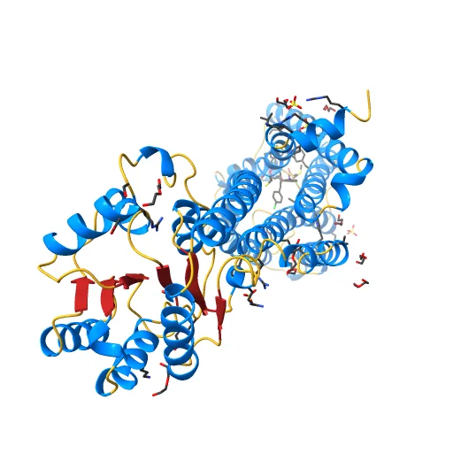 High-resolution crystal structure of the human CB1 cannabinoid receptor
 CRYSTAL STRUCTURE OF A SOLUBLE FORM OF THE HUMAN T CELL CO-RECEPTOR CD8 AT 2.6 ANGSTROMS RESOLUTION
 Crystal Structure of the extra-cellular domains of Human Interleukin-6 Receptor alpha chain
 Crystal Structure of GTP-bound ADP-ribosylation Factor Like Protein 1 (Arl1) and GRIP Domain of Golgin245 COMPLEX
 Crystal Structure of mouse Ryanodine Receptor 2 SPRY2 Domain (1080-1253)
 Crystal structure of Cas9 bound to PAM-containing DNA target
 Crystal structure of Rab8 in its inactive GDP-bound form
 Cytochrome C (Human) (PDB 1J3S)
 Protein Structure for PDB 3U8VThis is my second attempt to print out a protein structure based on a pdb file. This printed well, and is now a completed model. I am planning to paint it soon, and will post more later.
 Plastocyanin (pdb 2CJ3)This is a model of the structure of the electron transport protein plastocyanin. The model is based on the crystal structure and the PDB file 2CJ3. This printed well, and is now a completed model.
 FeMo-cofactor from NitrogenaseThis is a model of one of the most complicated metal clusters known in biology. I am preparing this and other cofactors and protein 3D Models.
 BCC Crystal StructureThis is a model of a BCC(Body-Centered Cubic) structure. I'm currently taking a materials engineering class so I decided to make a learning model. I plan on making 2 more, FCC & HCP. 40mm Sphere
 Flexible Perovskite Structure
 BCC, FCC AND HCP Unit Cell Structures
 Progesterone Receptor bound to DNA
 4M64: 3D crystal structure of Na+/melibiose symporter of Salmonella typhimurium
 PMCA3 Ca2+-Bound (modeled on SERCA structure 1SU4)
 High resolution crystal structure of the SMN Tudor domain
 Crystal structure of the Luciferase
 STRUCTURE OF T-CELL SURFACE GLYCOPROTEIN CD4, TETRAGONAL CRYSTAL FORM
 Crystal structure of the C-terminal Hexokinase domain of human HK3
 Crystal structure of E. coli decanoyl-ACP
 The Crystal Structure of Nucleoplasmin-Core
 X-RAY CRYSTAL STRUCTURE OF R111H HUMAN GALECTIN-1
 CRYSTAL STRUCTURE OF THE NEURONAL T-SNARE SYNTAXIN-1ADOI: 10.2210/pdb1EZ3/pdb Classification: ENDOCYTOSIS/EXOCYTOSIS Organism(s): Rattus norvegicus Expression System: Escherichia coli Deposited: 2000-05-09 Released: 2000-06-07 Deposition Author(s): Lerman, J.C. , Robblee, J. , Fairman, R.
 X-ray crystal structure of coil 1A of human vimentin
 Structure of the TRPM8 cold receptor by single particle electron cryo-microscopy, calcium-bound stateTRPM8 cold receptor, calcium-bound state
 Crystal structure of human wild-type GAPDH at 1.99 angstroms resolution
 Crystal structure of T. fusca Cas3
 Crystal Structure Of Biotin Protein Ligase From Aquifex Aeolicus
 CRYSTAL STRUCTURE OF THE EXPORTIN CSE1P COMPLEXED WITH ITS CARGO (KAP60P) AND RANGTP
 Crystal structure of the HPV16 E6/E6AP/p53 ternary complex at 2.25 A resolutionPDB 4XR8 Chain A
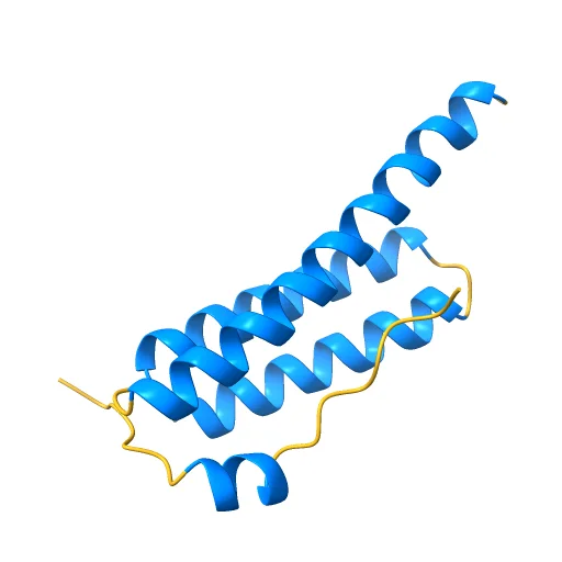 Crystal Structure of Vps4 MIT-Vfa1 MIM2
 CRYSTAL STRUCTURE OF RECOMBINANT HUMAN TRIOSEPHOSPHATE ISOMERASE AT 2.8 ANGSTROMS RESOLUTION. TRIOSEPHOSPHATE ISOMERASE RELATED HUMAN GENETIC DISORDERS AND COMPARISON WITH THE TRYPANOSOMAL ENZYME
SliSearch lists public model details and links to the original source. It does not host files or verify licences.