
NIH 3D
Crystal structure of (CENP-A/H4)2 heterotetramer


- Creator (as listed at the source)
- Not listed at the source.
- Licence
- Public DomainThe licence is listed by the source marketplace. SliSearch does not verify it.
- File types
- GLB STL
- Pricing
- Free
- Published
- Category
- Biomacromolecules
- Downloads
- 261
Overview
PDB Classifiation: DNA BINDING PROTEIN
Print this model with Slicience
- Download the model from the source page.
- Upload the file to Slicience and get a quote with Prinnu.
Similar models
Chosen by shared tags first, then category. Each one links to its own marketplace.
 CYP1A2 (Cytochrome P450 1A2)Imaged is my first printout of a PDB 2HI4 molecule cross-section (Cytochrome P450 1A2, or CYP1A2 for short, complex with alpha-napthoflavone ligand).
 Mycobacterium smegmatis Dps tetragonal formPDB Classifiation: DNA BINDING PROTEIN
 Protein secondary structureThese are teaching models of various protein secondary structures in space fill and stick representations (PDB: 3vjo .
 crystal structure of Se-substituted arabidopsis thaliana SHH1 SAWADEE domain L200M/L218M mutantPDB Classifiation: GENE REGULATION
 Peppytides: 3D printed, scaled, foldable model of the polypeptide chain for protein foldingFor a brief History of invention, Citation details and Contact details, please refer to Attribution. Peppytide is a scaled, flexible protein model to explore protein folding with hands.
 Structural basis of NR4A1 bound to the human pituitary proopiomelanocortin gene promoterPDB Classifiation: DNA BINDING PROTEIN/DNA
 Crystal structure of the LXRalfa-RXRbeta LBD heterodimerPDB Classifiation: DNA BINDING PROTEIN
 Cryptochrome 3 from Arabidopsis thalianaPDB Classifiation: DNA BINDING PROTEIN
 Structure of a glutamine-rich domain from histone deacetylase 4PDB Classifiation: TRANSCRIPTION
 Crystal structure of Rad4-Rad23 bound to a UV-damaged DNAPDB Classifiation: DNA BINDING PROTEIN/DNA
 Crystal structure of BioQPDB Classifiation: DNA BINDING PROTEIN
 AncMR DNA Binding Domain - (+)GRE ComplexPDB Classifiation: dna binding protein/dna
 Model for bacteriophage fd from cryo-EMPDB Classifiation: VIRUS
 355 bp di-nucleosome harboring cohesive DNA terminiPDB Classifiation: DNA BINDING PROTEIN
 Crystal structure of a Fragment of Nuclear factor related to kappa-B-binding protein (residues 370-495) (NFRKB) from Homo sapiens at 2.18 A resolutionPDB Classifiation: Transcription regulation, DNA BINDING
 Structures of inactive and activated DntR provide conclusive evidence for the mechanism of action of LysR transcription factorsPDB Classifiation: TRANSCRIPTION
 Alpha helixAlpha helix model, from the sequence Gly-Ala18-Gly. Constructed using Peptide Builder in Chimera. Includes backbone hydrogen bonds and side chains represented as sticks, as well as the "cardinal" backbone representation to more accurately...
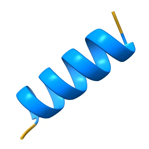 Pi Helix - Secondary StructureAn alpha helix is a commonly-found protein secondary structure. It is a right-handed coil in which every backbone N-H group donates a hydrogen bond to the C=O group of the amino acid four residues earlier.
 The structure of the centromeric nucleosome containing CENP-APDB Classifiation: STRUCTURAL PROTEIN/DNA
 Peppytides: a foldable model of the polypeptide chain
 Structure of Zeta-Zeta Transmembrane DimerPDB Classifiation: MEMBRANE PROTEIN
 Structure of TAL effector PthXo1 bound to its DNA targetPDB Classifiation: TRANSCRIPTION/DNA
 Crystal Structure of the heterotypic nucleosome containing human CENP-A and H3.3PDB Classifiation: DNA BINDING PROTEIN/DNA
 The First Fibronectin Type II Module from Human Matrix Metalloproteinase 2PDB Classifiation: HYDROLASE
 Solution structures of the C2H2 type zinc finger domain of human Transcriptional repressor CTCFPDB Classifiation: DNA BINDING PROTEIN
 Solution Structure of the SWI1 ARIDPDB Classifiation: DNA BINDING PROTEIN
 Helix variations - Secondary StructureA demonstration of the various alpha helix conformations in protein secondary structure. An alpha helix is a right-handed coil in which every backbone N-H group donates a hydrogen bond to the C=O group of the amino acid four residues...
 THE CRYSTAL STRUCTURE OF DPS, A FERRITIN HOMOLOG THAT BINDS AND PROTECTS DNAPDB Classifiation: DNA BINDING PROTEIN
 Cryo-EM structure of Lambda Phage protein GamS bound to RecBCD.PDB Classifiation: DNA BINDING PROTEIN
 Crystal structure of TREX1 in complex with a duplex DNA with 3' overhangPDB Classifiation: DNA BINDING PROTEIN
 TATA BINDING PROTEIN (TBP)/DNA COMPLEXPDB Classifiation: TRANSCRIPTION/DNA
 Crystal structure of two-domain RSV INTEGRASE covalently linked with DNAPDB Classifiation: DNA BINDING PROTEIN
 Crystal structure of 167 bp nucleosome bound to the globular domain of linker histone H5PDB Classifiation: CHROMATIN BINDING PROTEIN/DNA
 Structure of the M3 muscarinic acetylcholine receptor bound to the antagonist tiotropium crystallized with disulfide-stabilized T4 lysozyme (dsT4L)PDB Classifiation: MEMBRANE PROTEIN
 X-ray Structure at 1.75 A Resolution of a Norovirus Protease Linked to an Active Site Directed Peptide InhibitorPDB Classifiation: HYDROLASE/HYDROLASE INHIBITOR
 Structure of Human PARP-1 bound to a DNA double strand breakPDB Classifiation: TRANSFERASE/DNA
 Crystal structure of Dna2 nuclease-helicasePDB Classifiation: HYDROLASE
 Crystallization and structure determination of the core-binding domain of bacteriophage lambda integrasePDB Classifiation: DNA BINDING PROTEIN
 Structure of the Nuclear Factor Kappa-b (Nf-kb) P50 Homodimer
 Crystal structure of the hBAF250b AT-rich interaction domain (ARID)PDB Classifiation: DNA BINDING PROTEIN
SliSearch lists public model details and links to the original source. It does not host files or verify licences.